
Bacteria can gain novel traits, like antibiotic resistance, by sharing DNA with one another through processes cumulatively termed horizontal gene transfer.
One major mechanism of horizontal gene transfer is called natural transformation, a process by which a bacterium can take up free DNA from the environment and then integrate that DNA into its genome. Bacteria take up DNA from the environment using long hair-like surface appendages called pili (see “DNA-reeling bacteria yield new insight on how superbugs acquire drug resistance” and “IU scientists watch bacteria 'harpoon' DNA to speed their evolution”).
In a new study, published in the journal Cell, IU researchers focus on characterizing how bacteria can integrate this DNA into their genome after taking it up from the environment. This work was led by Ankur Dalia, an assistant professor in the Indiana University College of Arts and Sciences' Department of Biology.
Attempting to better understand DNA integration, the Dalia lab developed new methods to directly observe this process in real time in single bacterial cells under the microscope. Specifically, they developed a number of fluorescent "reporters" to tell exactly (1) when cells were trying to integrate DNA (see Video 1), (2) when cells succeeded at integrating this DNA, and (3) when the new DNA that they integrated was expressed during the process.
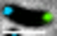 This video shows a Vibrio cholerae cell where two distant regions of the bacterial DNA genome are “painted” so that we can see them under a microscope—one area is marked with a blue protein, while the other is marked with a green protein. These cells also express a red protein fused to ComM, which is a protein that is required for the integration of DNA during natural transformation. In this video, these V. cholerae cells were given DNA that should integrate in close proximity of the blue painted region within the genome. Because we see that the red ComM protein goes to that location (and overlaps with the blue spot in the cell), this tells us that watching where the red ComM protein goes in the cell can tell us when these cells are trying to integrate DNA into their genome. This allowed us to determine how well cells can carry out this important process, which is critical for bacteria to acquire new traits like antibiotic resistance.
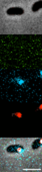 This video shows a Vibrio cholerae cell where two nearby regions of the bacterial DNA genome are “painted” so that we can see them under a microscope—one area is marked with a blue protein; the other is marked with a red protein. These cells were then given DNA that should delete the red marked region of the genome and introduce a gene that can code for a green protein. In this video, we first see that the cell integrates DNA into its genome (which is indicated by a loss of the bright red spot in the cell), it then replicates its genome (which is indicated by the “splitting” of the blue spot within the cell), and finally we see that the cell rapidly makes the green protein that is encoded by the newly acquired DNA. Interestingly, we see that when the blue spots “split” (aka, the genome is replicated), that the spot on the right also has a bright red spot. This means that the genome on the right did NOT acquire the new DNA and that only the genome on the left side of the cell (aka, the blue spot on the left) has the newly introduced DNA. This means that once this cell divides, only the cell on the left actually has the DNA to make the green protein. But at the end of the video, we can see that both the cell on the left and the cell on the right have acquired the green protein. This means that even though the cell on the right does not have the DNA to make the green protein, it can still acquire the green protein. This suggests that this bacterial process can mediate “nongenetic” inheritance, which was not previously appreciated.
 In this video, focus your attention on the cell indicated by the red arrow. Cells in this experiment were given DNA that would let them express two things: a green protein and a protein that provides resistance to an antibiotic. These experiments were images in the presence of the antibiotic, so if cells grow, it means that they are resistant to the drug. Once the indicated cell divides, you can see that the resulting cell on the left goes on to become bright green and continues to grow and divide. This is the cell that inherited the DNA that encodes the green protein and the antibiotic resistance gene. Go back to the beginning of the video and now watch the indicated cell divide again—if you look at the cell on the right, you will see that this cell ALSO continues to grow and divide but does not become bright green. This cell did not inherit the DNA that codes for the green protein OR the antibiotic resistance protein, BUT it can still grow in the presence of the antibiotic. This is because it inherited the antibiotic resistance protein WITHOUT inheriting the DNA that makes that protein. Compare the growth of this cell to cells that did not receive any DNA, which can be seen in the bottom right of the video. These cells do not grow in the presence of the antibiotic. This suggests that this bacterial process can mediate “nongenetic” inheritance, which was not previously appreciated.
 The College of Arts